
Guided Orthognathic Surgery: In-House 3D Printing and Patient-Specific Implants
Serge Ketoff · Gabrielle Magnant · Natacha Niez
CLINIC No. 452/453 · July–August 2025
Orthognathic surgery has benefited from digital tools: the creation of a digital clone of the patient makes it possible to plan the surgery on a computer — skeletal acquisition (CT scan or cone beam), dental acquisition (optical impression) and skin surface acquisition (photogrammetry), or even kinematic acquisition by recording joint range of motion.
From 2D Planning to the Digital Clone
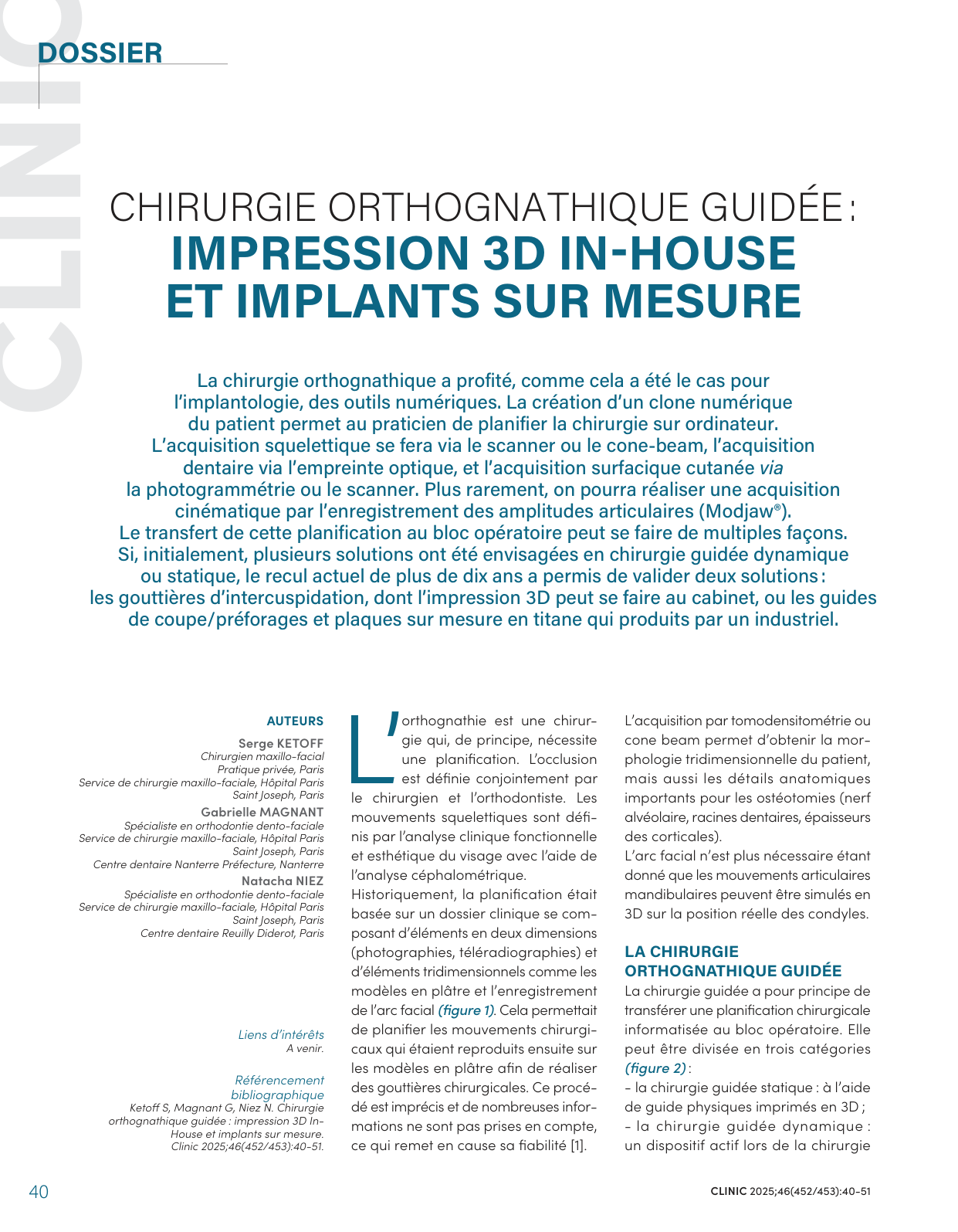
Historically, planning relied on photographs, cephalometric radiographs, plaster models and the facebow: "tracing-paper surgery", imprecise and incomplete. 3D imaging provides the patient's complete morphology and the anatomical details essential to osteotomies (alveolar nerve, tooth roots, cortical bone thickness). Three modalities of guided surgery exist — static (3D-printed guides), dynamic (navigation, augmented reality) and robotic — but only static guided surgery is used in routine practice.
Clinical Case: In-House Printed Occlusal Splints
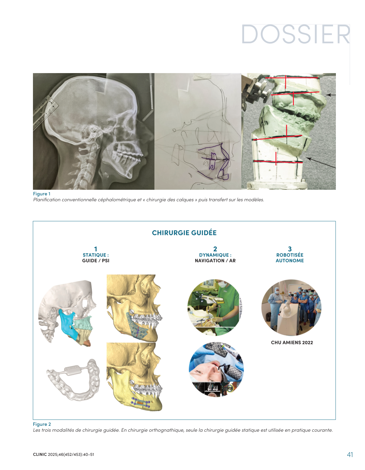
In a 17-year-old female patient with a skeletal Class II malocclusion and severe sleep apnea, 3D surgical planning precisely quantified a bimaxillary advancement with counterclockwise rotation of the maxilla. The software generated the intermediate and final occlusal splints, printed in the office on a stereolithographic printer. Superimposition of pre- and postoperative imaging demonstrated a significant increase in airway volume.
Clinical Case: Cutting Guides and Patient-Specific Titanium Plates
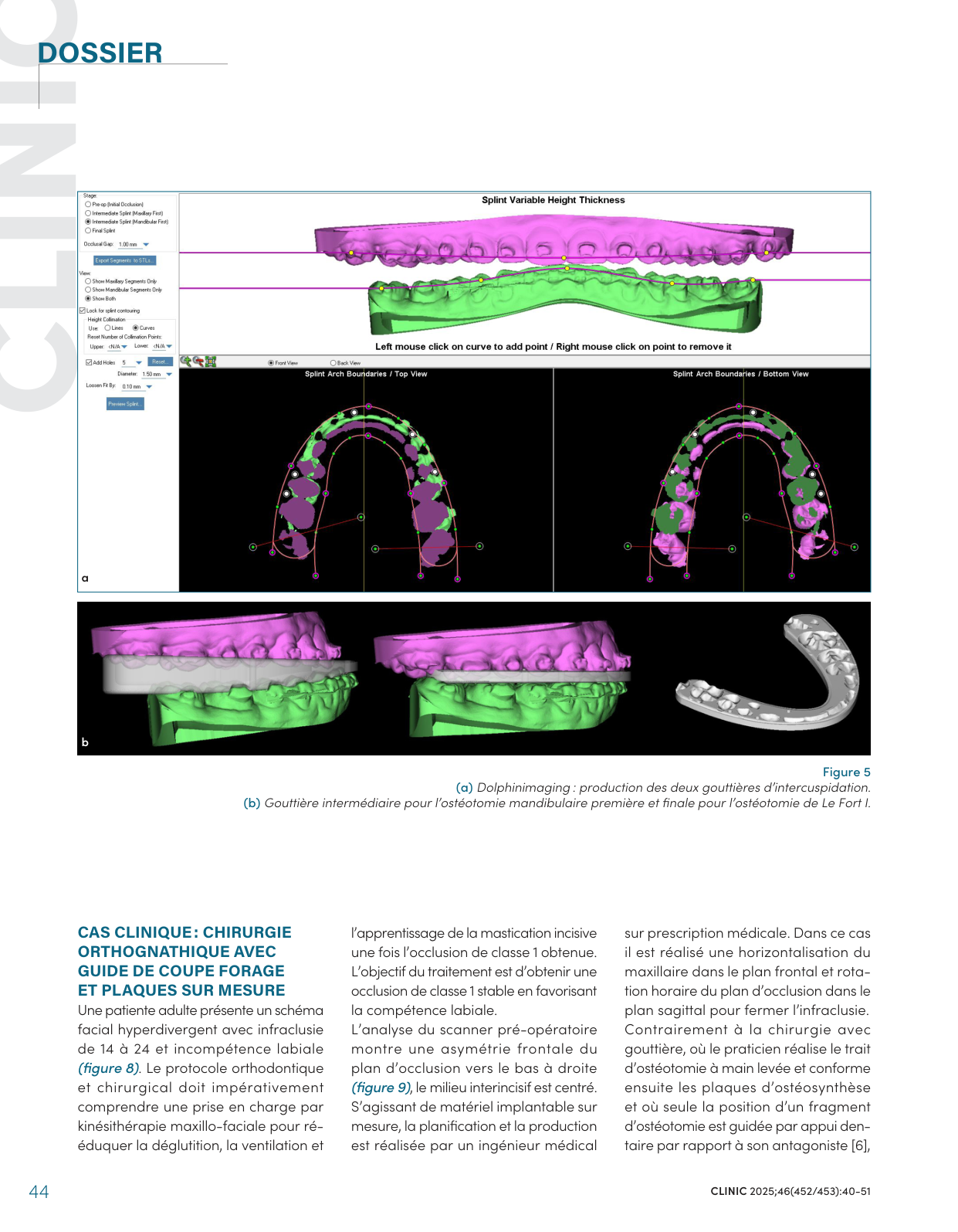
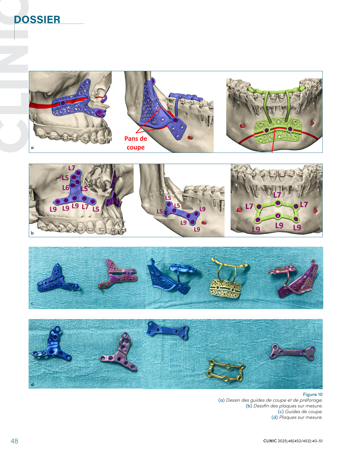
For a hyperdivergent female patient with anterior open bite and lip incompetence, planning was carried out with a biomedical engineer: cutting and predrilling guides, followed by patient-specific printed titanium plates that position the osteotomy segment on a bony reference, without intermaxillary fixation. This bone-based, rather than occlusion-based, repositioning is considered more accurate — however, the osteotomy design can no longer be adapted intraoperatively.
Reference: Ketoff S, Magnant G, Niez N. Chirurgie orthognathique guidée : impression 3D In-House et implants sur mesure [Guided orthognathic surgery: in-house 3D printing and patient-specific implants]. Clinic 2025;46(452/453):40-51.